Phacoemulsification removes the cloudy natural lens through a self-sealing opening that is only a few millimetres wide, then places an artificial lens inside the remaining capsule. By the end, you will understand each surgical step, what the small incision changes, which examinations determine suitability, and when another technique may be safer.
Key takeaways
- A 2–3 mm incision lets the surgeon remove the cloudy lens in small fragments.
- Biometry, corneal measurements and retinal checks help confirm whether phacoemulsification suits you.
- Dense cataracts, weak eye support or corneal disease can make small-incision surgery harder.
- Ask about the incision, intraocular lens, recovery restrictions and alternatives before consenting.
How the cloudy lens is removed through a 2–3 mm opening
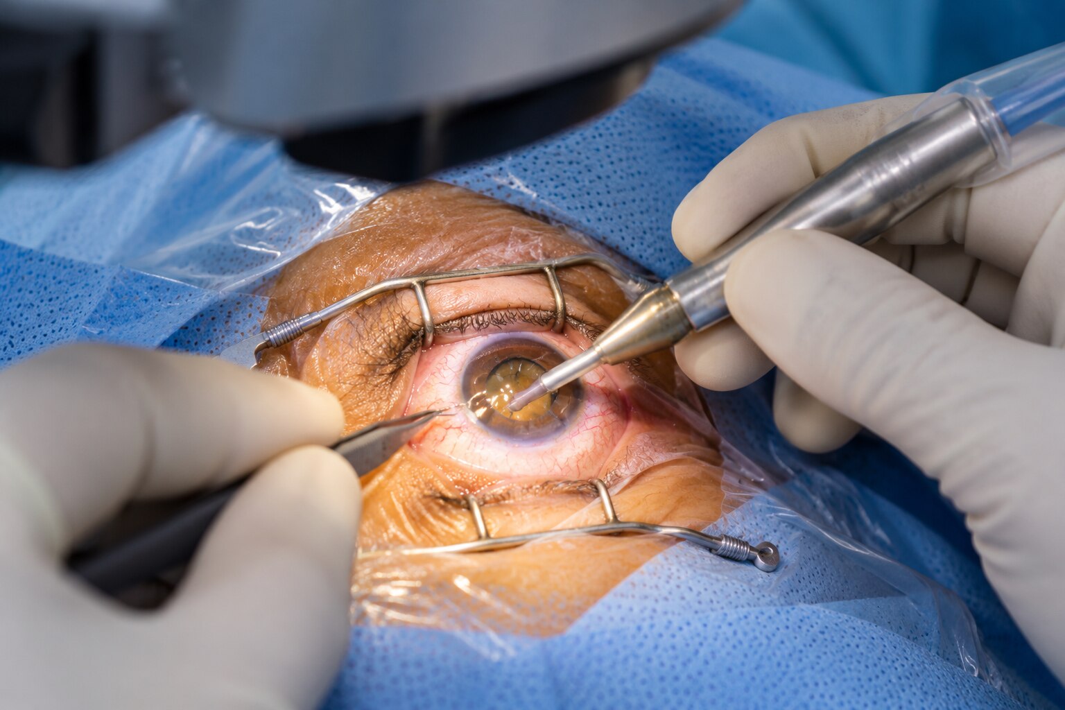
A 2–3 mm corneal incision gives the surgeon access to the cloudy natural lens without removing the surrounding eye tissue. This small incision cataract surgery approach uses a self-sealing wound design, although the exact width depends on the instruments, incision construction and intraocular lens.
The lens is removed in a controlled sequence:
- The surgeon makes the corneal incision and fills the front chamber with balanced fluid to maintain its shape.
- Through the opening, the surgeon creates a circular capsulorhexis in the front of the thin lens capsule.
- A phacoemulsification handpiece delivers ultrasound energy to break the firm, cloudy lens nucleus into small fragments. Irrigating fluid cools and protects the working area while aspiration draws the fragments out through the probe.
- The softer lens material is removed, while the posterior capsule is left in place to support the replacement lens.
- A folded intraocular lens is inserted through the same small opening and unfolded inside the remaining capsular bag.
The artificial lens restores the eye’s focusing power for distance or another selected target, but it does not reproduce the natural lens’s ability to change focus for near vision. Phacoemulsification surgery uses ultrasound, not a laser, to fragment the cataract; a femtosecond laser can assist selected steps but does not eliminate phacoemulsification.
The wound often heals without stitches because its narrow, angled construction closes under normal eye pressure.
What happens to your eye before, during and just after surgery
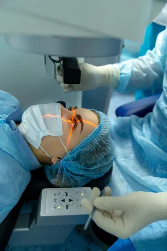
You are usually awake throughout the 15–30-minute operation, although the surgical unit visit takes longer. Local anaesthetic for cataract surgery numbs the eye; you may notice touch, pressure or bright lights, but sharp pain is not expected.
1. Before the procedure, dilating eye drops widen your pupil. The team cleans the skin around your eye, places a sterile drape, and checks your medicines and general health. You lie on your back and must keep your head still while breathing normally.
2. During surgery, the surgeon makes a tiny corneal opening and works through it with fine instruments. You may see movement or changing colours without being able to follow the details. A lid holder keeps you from blinking, and the operation usually feels more like pressure than pain.
3. Afterward, the eye is checked and covered with a shield or pad. Vision can be hazy at first, with watering, mild irritation, light sensitivity or temporary corneal swelling. Use the prescribed drops and avoid rubbing or pressing the eye during cataract surgery recovery.
The wound often needs no stitch because the surgeon shapes it as a short, angled, valved tunnel. Eye pressure helps close this self-sealing incision, reducing wound healing time and surgically induced astigmatism. A stitch is still needed if the opening is enlarged, unstable or leaking.
Increasing pain, marked redness, worsening vision, flashes or new floaters needs urgent assessment.
Which tests show whether phacoemulsification is suitable
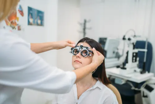
A cataract surgery evaluation combines an examination of eye health with measurements that predict surgical risk and calculate the replacement lens. The decision is not based on cloudiness alone: macular degeneration, glaucoma, diabetic retinopathy, or corneal disease can limit vision after the cataract is removed.
| Test | What it checks | How it guides the decision |
|---|---|---|
| Slit-lamp examination | Cornea, iris, pupil, lens, capsule and inflammation | Finds corneal scars, a dense cataract, weak supporting structures or active inflammation that could make surgery less safe |
| Eye pressure measurement | Intraocular pressure | Identifies glaucoma or pressure that needs treatment before or after surgery |
| Dilated retinal examination | Retina, optic nerve and macula | Detects disease that cataract surgery cannot reverse and may require additional testing |
| Biometry for intraocular lens power | Axial length, corneal curvature and sometimes anterior-chamber depth | Calculates the intraocular-lens power intended to produce the planned focusing result |
| Macular OCT or B-scan ultrasound | Macular detail or the retina behind an opaque lens | Reveals hidden retinal disease when the cataract blocks a clear view |
Biometry for intraocular lens power is most reliable when the eye surface is healthy and the measurements are repeatable. Dry eye, an unstable tear film, previous corneal surgery, or an unusually long or short eye can reduce accuracy, so the surgeon may treat the surface or repeat the scan.
A retinal finding can make phacoemulsification technically suitable while changing the expected visual improvement.
When a small-incision approach becomes more difficult
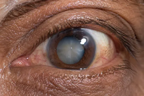
A hard cataract, weak zonules, a small pupil, or disease elsewhere can make the small-incision approach less predictable. Dense cataract surgery needs more mechanical work and ultrasound energy, which can stress corneal endothelial cells, cause postoperative corneal swelling, and delay clear vision.
Zonular weakness or a fragile lens capsule raises the risk of lens instability or a capsule tear.
| Problem | Why phacoemulsification becomes harder | Limitation afterward |
|---|---|---|
| Corneal disease or low endothelial-cell count | Ultrasound and fluid movement can worsen the cornea | Blurred vision and swelling may last longer; severe damage can limit recovery |
| Retinal disease, macular degeneration, or diabetic retinopathy | Removing the cataract cannot repair damaged retinal tissue | Vision may improve less than expected, even with a clear lens |
| Glaucoma or high eye pressure | Surgery and postoperative medicines can alter pressure | Pressure monitoring and additional treatment may be needed |
| Small pupil or capsule scarring | The surgeon has less room to see and manipulate the lens | Extra instruments, a longer procedure, or a different technique may be required |
| Poor positioning, tremor, or inability to lie flat | Eye movement and access become harder during surgery | The procedure may need additional support, anaesthesia planning, or postponement |
A very dense cataract or unstable capsule can require a larger opening or another cataract-removal technique instead of routine phacoemulsification. Femtosecond-laser assistance does not eliminate these problems or guarantee better vision. After surgery, expect possible inflammation, temporary blur, glasses, and slower recovery; sudden pain, marked redness, worsening vision, flashes, or floaters require urgent assessment.
Questions to settle before choosing your cataract procedure
Choose the lens and surgical method only after you know what vision you want, what your eyes can support, and what glasses you are willing to use afterward. Ask the specialist to compare these intraocular lens options:
| Option | What it means | What to clarify |
|---|---|---|
| Monofocal lens | Focuses at one chosen distance, often far | Which reading or computer glasses will you need? |
| Multifocal lens | Provides more than one focal range | Are halos, glare, reduced contrast, or night-driving problems acceptable? |
| Toric lens | Corrects corneal astigmatism as well as cataract-related blur | Is your astigmatism regular enough for this lens, and will it need precise alignment? |
- Ask what vision is realistic after surgery, including reading, driving at night, and dependence on glasses. Macular degeneration, glaucoma, diabetic retinopathy, or corneal disease can limit the result.
- Ask whether standard manual phacoemulsification suits you or whether femtosecond-laser assistance offers a specific benefit. It does not remove the need for phacoemulsification and can add cost.
- Ask which cataract surgery complications matter in your case, including infection inside the eye, retinal detachment, macular swelling, raised eye pressure, and lens-position problems.
- Confirm the eye-drop schedule, review dates, activity restrictions, emergency number, and who examines you if pain, marked redness, flashes, floaters, or worsening vision develop.
- At Focus Eye Clinic - 02235048608, ask which hospital, surgeon, anaesthesia team, emergency service, and backup arrangements support the operation.
Related services
Ophthalmology Phacoemulsification Consultation in Thane ₹900 Phacoemulsification Surgery is an advanced and safe cataract treatment available at Focus Eye Clinic by Dr. Mohini Modak. View service → |
Ophthalmology Cataract Consultation in Thane ₹900 Cataract Surgery at Focus Eye Clinic by Dr. Mohini Modak is a safe and effective procedure to restore clear vision affected by cataracts. View service → |
Frequently asked questions
How is the cloudy lens removed through a 2–3 mm opening?
The surgeon opens the lens capsule, uses ultrasound energy to break the cloudy lens into fragments, and gently removes them through the incision before placing an intraocular lens.
What happens before, during and just after phacoemulsification surgery?
Before surgery, the eye is measured and examined. During surgery, the eye is numbed and the cloudy lens is removed. Afterward, you receive eye drops and instructions for protecting the eye during recovery.
Which tests show whether phacoemulsification is suitable?
The assessment can include visual acuity, slit-lamp examination, eye-pressure measurement, corneal evaluation, biometry for intraocular lens power, and a retinal examination. Ultrasound may be needed when the cataract blocks the retina view.
When does a small-incision approach become more difficult?
Dense or mature cataracts, weak zonules, a shallow front chamber, corneal disease, poor pupil dilation or a limited view of the retina can make phacoemulsification more complex.
What should you ask before choosing a cataract procedure?
Ask which incision and intraocular lens are planned, whether your measurements are reliable, what risks apply to your eye, how long recovery takes, and what alternative procedure is available if phacoemulsification is unsuitable.
Keywords