A vision appointment can involve more than reading letters from a chart: different tests measure visual sharpness, focusing, eye coordination and ocular health. By the end, you will know what happens at each stage, which results are screening clues rather than diagnoses, how to prepare and when a routine test is not the right response to symptoms.
Key takeaways
- Bring your glasses, contact lens details, medicines and relevant eye history.
- Refraction measures your prescription; eye-health tests look beyond visual sharpness.
- Dilating drops can blur near vision and increase light sensitivity for several hours.
- Seek urgent care for sudden vision loss, severe eye pain or new flashes and floaters.
What happens when you first arrive for a vision assessment?
Your examination begins with a patient history: you describe blurred vision, headaches, eye strain, double vision, past eye conditions, medicines, injuries, surgeries and relevant family history. This helps the clinician choose tests and interpret your responses.
- Distance visual acuity measures how sharply you see letters or symbols on a chart from a specified distance, with each eye tested separately and then together.
- Near visual acuity measures your ability to read print at close range, often using a card held at a standard distance.
- Binocular vision testing checks how well your eyes work as a pair, including alignment, eye teaming, focusing and depth perception.
- The examiner records whether you use spectacles or contact lenses and compares performance with and without your current correction.
A good chart result does not prove that your eyes are healthy. Visual acuity measures central sharpness, but it does not assess peripheral vision, eye pressure, the optic nerve or the retina. Further examination is needed when symptoms, medical history or the initial findings suggest an eye-health problem.
How is your spectacle prescription measured and refined?
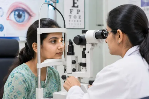
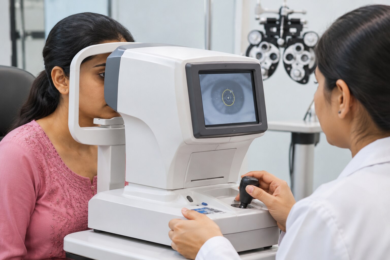
A visual-acuity chart shows how sharply you see; refraction measures the lens power that may improve that sharpness. The examiner begins with an objective estimate using an autorefractor, which analyses how light enters and leaves the eye.
Retinoscopy provides another objective method: the clinician shines a light into your eye and observes its movement through different lenses.
The estimate is refined during subjective refraction. You look at letters or symbols while the examiner changes lens powers, then compare options by saying which appears clearer. Your answers matter because the clearest lens is not always the strongest lens, and excessive power can cause strain or blur.
Accommodation—the eye’s ability to focus on near objects—can distort the result, especially in children, people with suspected latent long-sightedness, or those with accommodative spasm. Cycloplegic refraction uses drops to temporarily relax this focusing muscle before measurement. Without cycloplegia, active focusing can make the prescription appear too short-sighted.
An autorefractor reading is a starting measurement, not automatically your final prescription. The clinician checks it against retinoscopy, your responses, eye alignment, working distance and comfort. The finished prescription corrects vision; it does not by itself prove that your eyes are healthy.
Which tests check eye health beyond how clearly you see?
Good visual acuity does not rule out glaucoma, retinal disease or problems affecting peripheral vision. Eye-health testing looks beyond the letters you can read.
| Test | What it examines | What the result cannot establish |
|---|---|---|
| Colour vision | Ability to distinguish coloured dots, shapes or symbols | It does not assess sharpness, retinal health or the cause of an acquired colour change. |
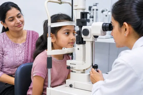
| Slit-lamp examination | Cornea, conjunctiva, iris, lens and other front-of-eye structures under magnification | A normal view of the front of the eye does not exclude retinal or optic-nerve disease. |
| Tonometry | Intraocular pressure; Goldmann applanation tonometry is a clinical reference method | One pressure reading neither confirms nor excludes glaucoma. Corneal thickness, biomechanics and time of day affect the result. |
| Visual-field testing | Sensitivity across central and peripheral vision while you respond to lights | A reliable field still needs comparison with the optic nerve, pressure and other findings. |
| Retinal assessment | Retina, blood vessels, macula and optic-nerve appearance through examination, sometimes after dilation | A normal appearance cannot detect every microscopic or early structural change. |
| OCT | Cross-sectional images of retinal layers and the optic-nerve fibre layer | A colour warning or thickness number is not a diagnosis; myopia, unusual discs, segmentation errors and media opacity can mislead. |
| Dry-eye evaluation | Symptoms, tear-film breakup time, surface staining, tear volume, eyelid margins and meibomian glands | One symptom or machine reading cannot define the whole condition. |
Clinicians interpret these results together. OCT findings need correlation with the examination and visual field, while dry-eye treatment depends on which tear-film or eyelid problem is present.
What should you expect if the clinician uses dilating drops?
Pupil dilation lets the clinician see a wider area of the retina and optic nerve than an undilated pupil allows. This improves a retinal examination and optic-nerve examination when checking for conditions such as retinal tears, macular disease or optic-nerve changes.
- Before the drops, tell the clinician about glaucoma, medication allergies, pregnancy, contact-lens use and any previous reaction to dilating drops. They may check your vision and eye pressure first.
- The dilating drops can sting briefly. Your pupils enlarge over about 15 to 30 minutes while you wait, and the clinician then examines the back of each eye with a light and lens.
- Afterward, near tasks such as reading a phone may look blurred, and bright light can feel uncomfortable. Wear sunglasses and avoid scheduling work that demands fine close vision immediately afterward.
Practical effects commonly last four to six hours, but blurred near vision or light sensitivity can continue into the evening, especially with stronger drops. Arrange someone else to drive if you cannot confirm that your vision is clear and comfortable; do not assume you can drive safely just because distance vision seems acceptable.
When is a screening, refraction or comprehensive examination appropriate?
Choose the least extensive option that answers your question, but do not use a clear chart result as proof that your eyes are healthy.
| Option | What it answers | When it applies |
|---|---|---|
| Vision screening | Whether vision or risk indicators need follow-up | School, workplace or community checks, or a quick first assessment |
| Spectacle power test | Which optical correction improves clarity | Blurred distance or near vision without known eye disease symptoms |
| Comprehensive ophthalmic examination | Whether an eye condition is present, and how it should be managed | Eye pain, recurrent redness, flashes, floaters, diabetes, glaucoma risk, abnormal screening, or a need for medical review |
An optometrist-led refraction is sufficient when your main problem is changed clarity and your history and eye-health checks show no warning signs. Refraction measures an optical prescription, not retinal health, eye pressure or the optic nerve; an autorefractor reading still needs clinical refinement.
Arrange a comprehensive ophthalmic examination with an ophthalmologist when symptoms, medical conditions, family history or an abnormal result require diagnosis. Focus Eye Clinic - 02235048608 can help arrange diagnostic review when a spectacle power test does not explain your symptoms.
Do not wait for routine testing after sudden flashes, a shower of new floaters, or a curtain-like shadow; seek urgent eye assessment because these symptoms can signal retinal tear or detachment.
How should you prepare, and when should you seek urgent eye care?
Bring your current spectacles, contact lenses, medication list, previous eye reports and relevant medical history. Record when symptoms began, whether one or both eyes are affected, and any diabetes, hypertension or previous eye surgery.
- Tell the examiner how long you wear contact lenses each day and when you last removed them. Bring the lenses and case if asked.
- Take sunglasses if pupil-dilating drops might be used; arrange help with driving because near vision blur and light sensitivity can last several hours.
- Seek urgent eye assessment for sudden vision loss, new flashes, a sudden shower of floaters, or a curtain or shadow across your vision. These symptoms can indicate a retinal tear or retinal detachment.
- Do not wait for a routine appointment when these symptoms appear. Contact an ophthalmic service promptly, especially if they affect one eye or follow an injury.
Routine blur, eye strain or a gradual prescription change can be assessed through a scheduled examination.
Frequently asked questions
What happens when you first arrive for a vision assessment?
You provide a patient history covering blurred vision, headaches, eye strain, double vision, eye conditions, medicines, injuries, surgeries and family history. The clinician uses this information to select and interpret tests.
How is a spectacle prescription measured and refined?
The clinician measures your current vision and tests different lens powers. You compare options during refraction so the prescription balances clarity, comfort and everyday visual needs.
Which tests check eye health beyond how clearly you see?
Eye-health testing can examine the front of the eye, eye pressure, pupil responses, eye movements and the retina or optic nerve. These checks can identify problems that do not reduce clarity at first.
What should you expect if the clinician uses dilating drops?
Dilating drops enlarge your pupils so the clinician can inspect more of the retina and optic nerve. Expect temporary blurred near vision and increased light sensitivity; bring sunglasses and avoid driving until your vision is comfortable and clear.
When is a screening, refraction or comprehensive examination appropriate?
A screening is a brief check for possible problems, refraction focuses on spectacle power, and a comprehensive examination assesses vision and eye health together. Choose a comprehensive examination for symptoms, eye disease risk or a longer gap since your last full assessment.