A clear result on a distance eye chart does not prove that every part of your eyes is healthy. A routine appointment can uncover focusing problems, cataract, glaucoma risk, retinal changes, corneal disease and conditions that cause few or no early symptoms; knowing which tests identify them helps you decide what follow-up you need.
Key takeaways
- A complete exam checks eye health, not just your spectacle prescription.
- Ask about focusing, tear-film, eyelid, corneal and retinal findings.
- Cataract, glaucoma and ocular hypertension are different conditions requiring different assessments.
- Seek prompt care for sudden vision loss, flashes, floaters or eye pain.
What a complete eye examination checks beyond spectacle power
A routine vision test usually measures how clearly you see at distance, often to decide whether your spectacle prescription needs changing. A complete eye examination checks eye health as well as sharpness, because normal visual-acuity testing does not rule out early disease.
During the appointment, the clinician may:
- Ask about blur, headaches, eye pain, flashes, medical conditions, medicines and family history.
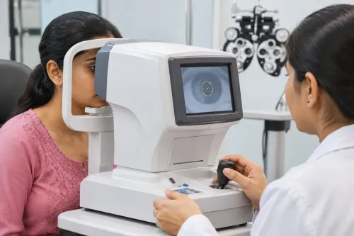
- Perform distance and near visual-acuity testing, followed by refraction to measure focusing error and presbyopia.
- Carry out pupil assessment and eye-movement testing to identify unequal pupils, nerve problems, or poor alignment.
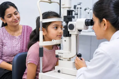
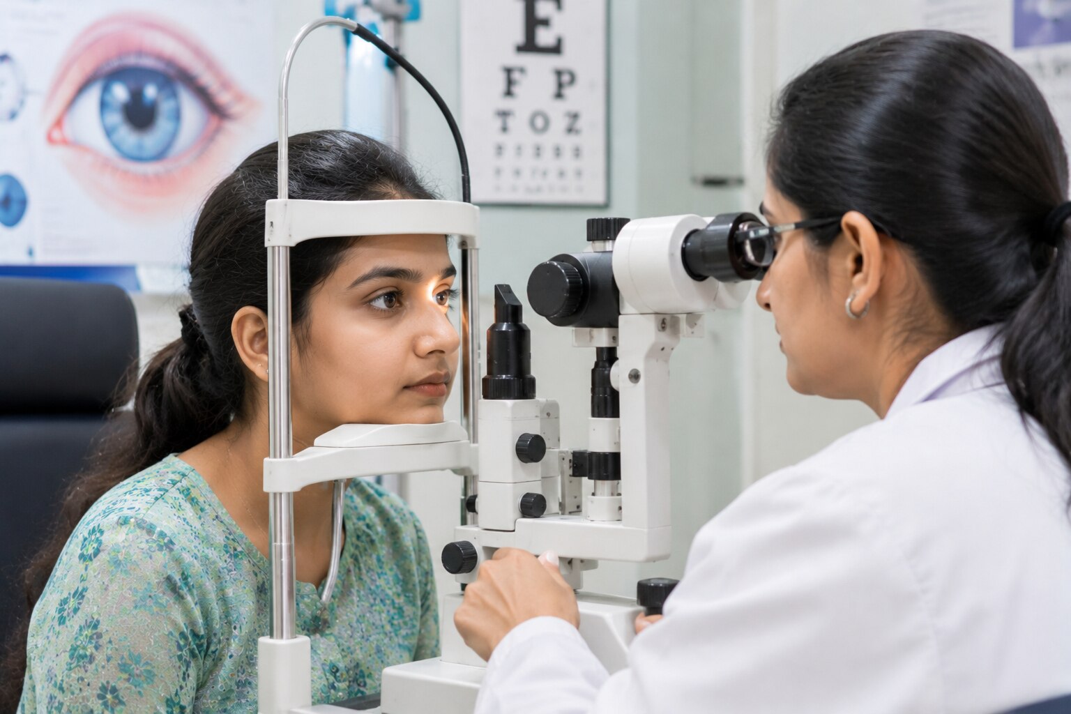
- Examine the eyelids, cornea, conjunctiva, lens and front of the eye with a slit lamp.
- Measure intraocular pressure with tonometry. A raised result alone does not diagnose glaucoma, and glaucoma can occur at statistically normal pressure.
- Test peripheral vision when glaucoma or another neurological concern is suspected.
- Examine the optic nerve and retina, often after dilation; gonioscopy assesses the drainage angle when indicated.
The extra checks can reveal cataract, dry-eye surface changes, keratoconus, glaucoma or diabetic retinal changes before you notice symptoms. Frequent prescription changes, irregular astigmatism or unexpectedly reduced best-corrected vision may prompt corneal topography or tomography.
A complete examination therefore takes longer than reading an eye chart, but it can detect problems that stronger glasses cannot correct.
Focusing, tear-film, eyelid and corneal conditions commonly found
An examination can identify refractive errors, tear-film instability, eyelid disease and corneal changes even when your vision seems clear. Refraction detects myopia, hyperopia, astigmatism and presbyopia; an eye-chart result alone cannot determine the correct prescription.
| Condition | What examination may show | Symptoms you may notice |
|---|---|---|
| Myopia, hyperopia, astigmatism or presbyopia | Reduced unaided acuity or a measurable refractive error | Blurred distance or near vision, eyestrain, headaches or difficulty reading |
| Dry-eye disease | Rapid tear-film breakup, reduced tear production or punctate corneal staining | Burning, grittiness, watering, fluctuating vision or light sensitivity |
| Blepharitis or meibomian-gland dysfunction | Crusted lashes, inflamed lid margins or blocked oil glands | Itching, sore lids, morning debris or intermittent blur |
| Corneal disease, including keratoconus | Irregular astigmatism, frequent prescription changes or reduced best-corrected vision | Distorted vision, glare, halos or increasing difficulty with glasses |
| Conjunctivitis | Redness and discharge on examination | Red eye, irritation, watering or sticky lids |
A slit-lamp examination assesses the eyelids, tear film and cornea directly. If refraction changes repeatedly or corrected vision remains unexpectedly poor, corneal topography or tomography can detect keratoconus more reliably than a stronger spectacle prescription. Good visual acuity does not rule out dry-eye damage, and limited discomfort does not prove the surface is healthy.
How cataract, glaucoma and ocular hypertension are distinguished
Good distance vision does not rule out eye disease, and a poor eye-chart result does not identify its cause. Clinicians combine unaided and corrected visual acuity with refraction, a slit-lamp examination, intraocular-pressure measurement and optic-nerve assessment rather than relying on one reading.
| Finding | Main evidence | What separates it |
|---|---|---|
| Refractive error | Myopia, hyperopia or astigmatism improves with an objective or subjective refraction | The eye is otherwise healthy, and suitable lenses usually improve clarity |
| Cataract | The slit-lamp examination shows clouding of the lens | Glare, halos, reduced contrast or changing prescriptions result from lens opacity; surgery depends on functional difficulty and examination findings, not an acuity number alone |
| Ocular hypertension | Intraocular pressure is repeatedly above the expected range | Pressure is raised without established optic-nerve damage or corresponding visual-field loss |
| Glaucoma | Optic-nerve damage, matching visual-field loss or both | Pressure may be high or fall within the statistically normal range; diagnosis depends on the pattern of structural and functional damage |
A clinician assessing possible glaucoma may add optical-nerve imaging, visual-field testing, gonioscopy to inspect the drainage angle and repeat pressure measurements. Ocular hypertension needs monitoring because it increases glaucoma risk, but treating the pressure is not the same as diagnosing glaucoma.
A cataract can be visible before major blur appears, while glaucoma can progress without pain or noticeable vision loss.
Retinal and macular findings that may appear before vision changes
A clear distance-vision result does not clear the retina. A dilated retinal examination can reveal disease before you notice blur, distortion or pain.
1. Diabetic retinopathy may appear as tiny microaneurysms, retinal haemorrhages or damaged retinal blood vessels. Macular oedema and proliferative changes carry greater risk to sight, yet early disease can coexist with normal visual acuity. An unexpected retinal finding does not diagnose diabetes; share it with your primary-care clinician.
2. Hypertensive retinal changes include narrowed or abnormal-looking vessels, bleeding and swelling near the optic disc. These signs can indicate the effects of high blood pressure on the eyes, but they do not confirm hypertension on their own. Arrange medical assessment when the finding is unexpected.
3. Age-related macular degeneration is assessed at the macula, the area responsible for detailed central vision. Drusen, pigment changes, fluid or bleeding can signal AMD before symptoms begin. AMD usually affects central rather than isolated peripheral vision.
4. Posterior vitreous detachment occurs when the gel inside the eye separates from the retina, often with ageing. It is not the same as retinal detachment, but new flashes, a sudden increase in floaters or a curtain-like shadow need prompt retinal evaluation because a retinal tear can accompany the separation.
The examination may also identify optic-disc abnormalities or other vascular changes before they affect everyday sight.
Who needs regular examinations and when a finding needs prompt care
Children need an eye examination at about 6 months, 3 years and before starting school; schedule earlier review if there is a family history of eye disease, poor visual behaviour or an abnormal screening result. A squint needs assessment because untreated misalignment can cause amblyopia, or reduced vision in one eye.
For routine review, use these intervals as a starting point:
- Healthy adults aged 20–39: every 5–10 years.
- Adults aged 40–64: every 2–4 years, with more frequent checks if symptoms or risk factors appear.
- Adults aged 65 and older: every 1–2 years.
- Contact-lens wearers: at least annually, or at the interval set by the clinician who checks the lenses and cornea.
- People with diabetes, glaucoma risk, previous eye disease or prescribed medicines that affect the eyes: follow the examination schedule set by their ophthalmologist; diabetes commonly requires a dilated retinal review at least yearly.
Seek same-day medical review for sudden vision loss, a curtain-like shadow, new flashes, or a sudden shower of floaters. An attack of severe eye pain with redness, nausea, vomiting or halos is also urgent. Pain, light sensitivity and reduced vision after a contact lens stays in require prompt corneal assessment.
Unexpected retinal bleeding, optic-disc change or vessel abnormality needs communication with your primary-care clinician; it does not diagnose diabetes or hypertension by itself. Focus Eye Clinic - 02235048608 can help determine whether a finding needs routine monitoring, testing or urgent referral.
Related services
Ophthalmology Eye Spectacles check up at Thane ₹900 Experience clarity and comfort with a comprehensive eye number check-up at Focus Eye Clinic, Thane. Led by Dr. View service → |
Ophthalmology ₹900 Retinal check-up at Focus Eye Clinic by Dr. Mohini Modak is essential for early detection of serious eye conditions that can affect vision. View service → |
Frequently asked questions
What does a complete eye examination check beyond spectacle power?
It checks eye health as well as visual sharpness, including the front of the eye, eye pressure and the retina.
Which focusing, tear-film, eyelid and corneal conditions can an examination find?
An examination can identify focusing problems, dry-eye or tear-film disruption, eyelid abnormalities and corneal changes that affect comfort or vision.
How are cataract, glaucoma and ocular hypertension distinguished?
Cataract affects the eye's lens. Glaucoma involves optic-nerve damage and corresponding visual-field loss. Ocular hypertension means raised eye pressure without established glaucomatous damage.
Can retinal or macular problems appear before vision changes?
Yes. Retinal and macular findings can be detected during examination before you notice a change in vision.
Who needs regular eye examinations?
People with diabetes, glaucoma risk, a family history of eye disease, changing vision or other eye concerns need a schedule advised by an eye-care professional.
Related services
Ophthalmology ₹900 Gonioscopy is a specialized diagnostic procedure used to examine the drainage angle of the eye, where the iris meets the cornea. View service → |
Ophthalmology ₹900 Dry Eye is a common eye condition that causes irritation, burning sensation, redness, watery eyes, and blurred vision due to inadequate tear... View service → |
Ophthalmology ₹900 At Focus Eye Clinic in Thane, Dr. Mohini Modak provides specialized care for uveitis, an inflammatory condition that can lead to permanent vision... View service → |
Ophthalmology ₹900 Amblyopia, commonly known as lazy eye, requires timely and specialized care to restore optimal vision. View service → |