Sharp distance vision does not prove that the macula, retinal blood vessels, peripheral retina or optic nerve are healthy. By understanding what a retinal check-up includes, when symptoms need urgent attention and how risk changes the review interval, you can choose the right examination instead of relying on a spectacle-number test alone.
Key takeaways
- Clear central vision does not rule out early retinal disease.
- Retinal check-ups can reveal damage before noticeable vision loss.
- Seek urgent assessment for sudden floaters, flashes, or a curtain-like shadow.
- Diabetes, high blood pressure, and family history can shorten screening intervals.
Why Clear Vision Can Miss Early Retinal Disease
A retina can deteriorate while central vision remains sharp. Early diabetic retinopathy, blood-vessel changes, macular disease or peripheral retinal damage may not affect the small central area used for reading and recognising faces. By the time blur appears, the disease may already be advanced.
A routine eye test or spectacle number test mainly checks how clearly you see and whether lenses improve that clarity. It does not reliably rule out hidden retinal disease.
| Test | What it checks | What it can miss |
|---|---|---|
| Routine eye test | Distance and near visual acuity | Retinal changes that have not reduced sharpness |
| Spectacle number test | Refraction and the lens strength needed for clearer focus | Disease behind the lens, including early retinal damage |
| Retinal examination | Retina, macula, retinal blood vessels and optic nerve, often after pupil dilation | Some conditions outside its scope, such as a complete glaucoma assessment |
A retinal examination lets the clinician inspect tissue that a clear letter chart cannot show. It can reveal bleeding, swelling, pigment changes or abnormal vessels before you notice symptoms, including changes linked to diabetes or high blood pressure.
An eye finding does not prove the cause or severity of a systemic illness, so medical follow-up may be needed.
Sharp vision is not permanent clearance. Retinal disease can develop after a normal visit, so your review interval depends on factors such as diabetes, high myopia, family history, previous eye disease, medicines and examination findings.
What Happens During a Retinal Check-Up
The appointment usually starts with history taking: tell the clinician about diabetes, high blood pressure, pregnancy, previous eye disease, medicines, family history and changes in vision. This information helps target the examination and identify risks that a spectacle-number test would miss.
- Visual acuity measures how sharply you read letters at different distances. It records central sight but cannot rule out early retinal damage when your vision still seems normal.
- Pupil examination checks how each pupil reacts to light and whether the responses are equal. An unusual response can point to problems involving the optic nerve or visual pathway.
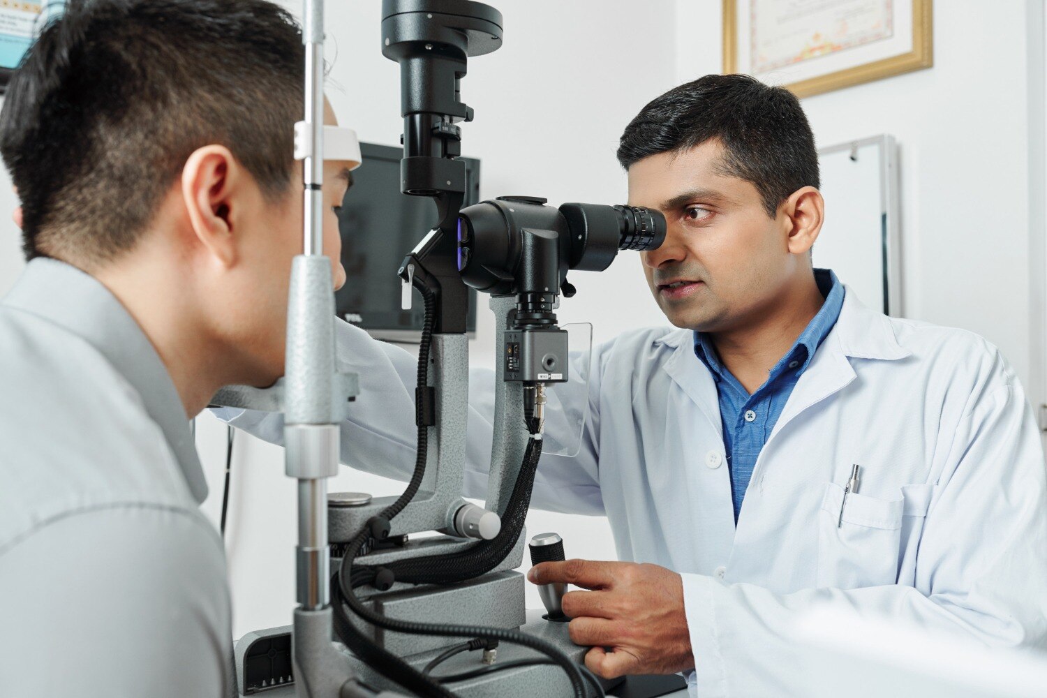
- A dilated fundus examination uses drops to widen the pupils, allowing inspection of the retina, macula, blood vessels and optic nerve. The clinician looks for bleeding, swelling, abnormal new vessels, pigment changes and structural damage.
- Retinal photography creates a recorded image of the back of the eye. Comparing photographs over time can reveal changes in vessels, the optic nerve or retinal lesions.
- Optical coherence tomography (OCT) produces cross-sectional retinal images. It can show fluid, swelling, thinning or subtle layer changes that are difficult to see directly.
- Fluorescein angiography may be considered when blood-vessel leakage or blockage needs closer assessment. After fluorescein dye enters a vein, timed photographs show how it moves through retinal vessels.
A retinal check is not a complete glaucoma assessment. Eye pressure, corneal thickness, optic-nerve appearance and visual-field testing may also be needed.
When Eye Symptoms Need Urgent Assessment
Do not wait for a routine appointment if you develop new flashes, a sudden increase in floaters, or a curtain over vision. These signs can indicate a retinal tear or detachment, which can permanently damage sight without urgent treatment.
Seek immediate assessment for sudden blurred vision, missing areas of vision, distortion of straight lines, or sudden loss of vision. A painful red eye, severe headache with visual changes, nausea, or halos around lights also needs prompt attention because high eye pressure, inflammation, corneal disease, or a neurological problem can be responsible.
- New flashes of light
- A sudden shower or increase of floaters
- A curtain or shadow across your field of vision
- Sudden blurred or lost vision
- A new blind spot or warped straight lines
A retinal check is valuable, but it is not a complete test for every eye complaint.
| Examination | Main target | What it cannot rule out |
|---|---|---|
| Retinal examination | Retina, macula, retinal blood vessels and optic nerve appearance | Every cause of pain, pressure-related damage, corneal disease or neurological vision loss |
| Glaucoma assessment | Eye pressure, optic nerve, cornea and visual field | A retinal check alone cannot confirm or exclude glaucoma |
| Routine sight test | Distance vision and spectacle prescription | Early retinal disease, peripheral loss or hidden macular damage |
Glaucoma can develop without pain, with peripheral vision disappearing first. Arrange urgent assessment for warning symptoms even if your last retinal examination was normal. A normal result describes that visit, not permanent protection from new disease.
How Your Risk Changes the Screening Interval
A normal result is a snapshot, not permanent clearance. Arrange the next review according to your risk rather than waiting for blurred vision: early retinal disease and glaucoma can remain symptom-free.
| Group | Starting point | Review interval |
|---|---|---|
| Low-risk adult | No diabetes, high myopia, family history or previous eye disease | Every 1–2 years, or the interval your clinician sets |
| Type 2 diabetes | Diabetes eye screening soon after diagnosis | Usually annually; findings and glucose control can shorten or extend this |
| Type 1 diabetes | Within five years of diagnosis | Usually annually after the first examination |
| High-risk patient | High myopia, strong family history, previous retinal disease or relevant medication | At least annually, with shorter reviews when advised |
| Pre-existing diabetes and pregnancy | Before pregnancy or during the first trimester | During pregnancy and after delivery according to the eye specialist’s plan |
| Pregnancy-related diabetes without pre-existing diabetes | Ask the maternity and eye teams whether additional screening is needed | Follow their specific advice |
Book an earlier appointment after a diabetes diagnosis, worsening glucose control, newly diagnosed or poorly controlled high blood pressure, pregnancy, a new retinal finding, or starting medication that can affect the retina, such as hydroxychloroquine.
A retinal finding linked with high blood pressure does not prove how severe the systemic disease is, so arrange medical follow-up too. New flashes, a sudden increase in floaters, a curtain-like shadow or sudden vision loss needs urgent assessment, not a routine review.
How to Prepare for and Compare a Retinal Check-Up
Before a dilated examination, arrange transport or plan not to drive until your vision clears. Dilating eye drops can cause light sensitivity and blurred near vision for several hours, so bring sunglasses, your medication list, diabetes records and previous eye reports.
- Tell the clinician if you are pregnant, have diabetes, glaucoma, medication allergies or a history of retinal treatment.
- Ask how long the appointment will take and whether you need someone to accompany you.
- Do not skip prescribed medicines unless the clinic specifically tells you to.
- Carry your current glasses, contact-lens details and a list of recent symptoms.
Compare clinics on practical details rather than on the word “screening” alone. Focus Eye Clinic - 02235048608 is one example to ask whether the appointment includes a dilated fundus examination and which tests are available on the day.
| Option | What it means | When it matters |
|---|---|---|
| Clinical examination | The clinician inspects the retina, macula, vessels and optic nerve after dilation | Essential even when imaging is offered |
| Retinal photographs | Permanent images for documentation and comparison | Useful for monitoring visible changes |
| OCT scan | Cross-sectional images that show fluid, swelling or thinning | Important for macular and structural changes |
| Fluorescein angiography | Dye-based images showing retinal blood-flow leakage or blockage | Used when ordinary examination and imaging leave a specific question |
| Follow-up plan | A stated review date, results process and referral route | Prevents an abnormal finding from being left unexplained |
Related service
Ophthalmology ₹900 Retinal check-up at Focus Eye Clinic by Dr. Mohini Modak is essential for early detection of serious eye conditions that can affect vision. View service → |
Frequently asked questions
Why can retinal disease develop without blurred vision?
Early disease can affect peripheral or microscopic retinal areas while the central retina still supports clear reading and face recognition.
What happens during a retinal check-up?
An eye specialist checks visual function and examines the retina, often using dilated examination and retinal imaging when indicated.
Which eye symptoms need urgent assessment?
Seek urgent assessment for sudden flashes, a sudden increase in floaters, a curtain-like shadow, sudden vision loss, or new distortion.
How does health risk affect retinal screening intervals?
Diabetes, high blood pressure, previous retinal disease, high myopia, and family history can require more frequent examinations.
Keywords